Cardiac Applications
Intraprocedure visualization of the esophagus using interventional C-arm CT as guidance for left atrial radiofrequency ablation.
Tognolini A, Al-Ahmad A, Wang PJ, Hsia HH, Herfkens RJ, Girard E, Moore T, Fahrig R.
Department of Radiology, The Lucas Center for MR Spectroscopy and Imaging, Palo Alto, CA 94304, USA.
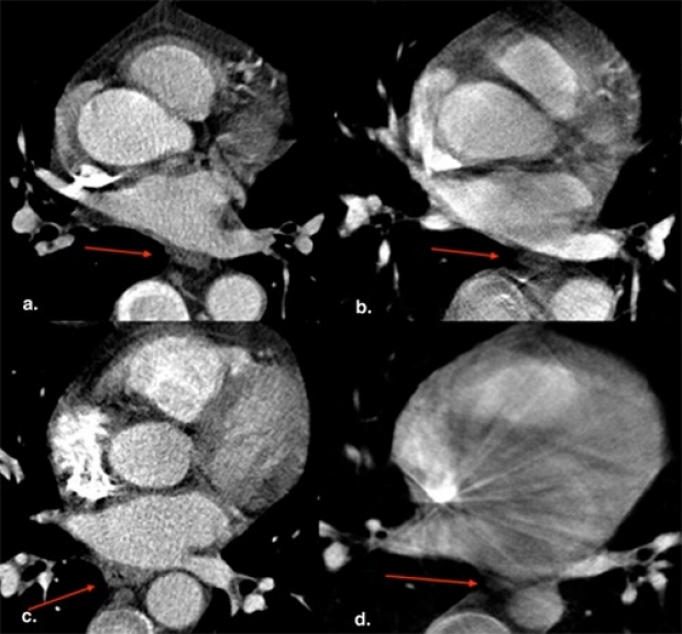
RATIONALE AND OBJECTIVES:: During radiofrequency catheter ablation for atrial fibrillation, the esophagus is at risk for thermal injury. In this study, C-arm computed tomography (CT) was compared to clinical CT, without the administration of oral contrast, to visualize the esophagus and its relationship to the left atrium and the ostia of the pulmonary veins (PVs) during the radiofrequency ablation procedure.
MATERIALS AND METHODS: Sixteen subjects underwent both cardiac clinical CT and C-arm CT. Computed tomographic scans were performed on a multidetector scanner using a standard electrocardiographically gated protocol. C-arm computed tomographic scans were obtained using either a multisweep protocol with retrospective electrocardiographic gating or a non-gated single-sweep protocol. C-arm and clinical computed tomographic scans were analyzed in a random order and then compared for the following criteria: (1) visualization of the esophagus (yes or no), (2) relationship of esophageal position to the four PVs, and (3) direct contact or absence of a fat pad between the esophagus and the PV antrum.
RESULTS:The esophagus was identified in all C-arm and clinical computed tomographic scans. In four cases, orthogonal planes were needed on C-arm CT (inferior PV level). In six patients, the esophageal location on C-arm CT was different from that on CT. Direct contact was reported in 19 of 64 of the segments (30%) examined on CT and in 26 of 64 (41%) on C-arm CT. In five of 64 segments (8%), C-arm CT overestimated a direct contact of the esophagus to the left atrium.
CONCLUSIONS:C-arm computed tomographic image quality without the administration of oral contrast agents was shown to be sufficient for visualization of the esophagus location during a radiofrequency catheter ablation procedure for atrial fibrillation.