Body Applications
In-vivo imaging of femoral artery nitinol stents for deformation analysis.
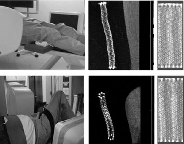
PURPOSE: The authors have developed a direct method to study femoral artery stent deformations in vivo. A previously described imaging and analysis approach based on a calibrated phantom was used to examine stents in human volunteers treated for atherosclerotic disease. In this pilot study, forces on stents were evaluated under different in-vivo flexion conditions.
MATERIALS AND METHODS: The optimized imaging protocol for imaging with a C-arm computed tomography system was first verified in an in-vivo porcine stent model. Human data were obtained by imaging 13 consenting volunteers with stents in femoral vessels. The affected leg was imaged in straight and bent positions to observe stent deformations. Semiautomatic software was used to calculate the changes in bending, extension, and torsion on the stents for the two positions.
RESULTS: For the human studies, tension and bending calculation were successful. Bending was found to compress stent lengths by 4% ± 3% (-14.2 to 1.5 mm), increase their average eccentricity by 10% ± 9% (0.12 to -0.16), and change their mean curvature by 27% ± 22% (0 to -0.005 mm(-1)). Stents with the greatest change in eccentricity and curvature were located behind the knee or in the pelvis. Torsion calculations were difficult because the stents were untethered and are symmetric. In addition, multiple locations in each stent underwent torsional deformations.
CONCLUSIONS: The imaging and analysis approach developed based on calibrated in vitro measurements was extended to in-vivo data. Bending and tension forces were successfully evaluated in this pilot study.
Imaging guidance with C-arm CT: prospective evaluation of its impact on patient radiation exposure during transhepatic arterial chemoembolization.

Ganguly A, Simons J, Schneider A, Keck B, Bennett NR, Herfkens RJ, Coogan SM, Fahrig R.
Department of Radiology, Stanford University, 1201 Welch Rd., Palo Alto, CA 94305, USA.
PURPOSE: To prospectively evaluate the impact of C-arm CT on radiation exposure to hepatocellular carcinoma (HCC) patients treated by chemoembolization.
MATERIALS AND METHODS: Patients with HCC (N = 87) underwent digital subtraction angiography (DSA; control group) or combined C-arm CT/DSA (test group) for chemoembolization. Dose-area product (DAP) and cumulative dose (CD) were measured for guidance and treatment verification. Contrast agent volume and C-arm CT utility were also measured.
RESULTS:The marginal DAP increase in the test group was offset by a substantial (50%) decrease in CD from DSA. Use of C-arm CT allowed reduction of DAP and CD from DSA imaging (P = .007 and P = .017). Experienced operators were more efficient in substituting C-arm CT for DSA, resulting in a negligible increase (7.5%) in total DAP for guidance, compared with an increase of 34% for all operators (P = .03). For treatment verification, DAP from C-arm CT exceeded that from DSA, approaching that of conventional CT. The test group used less contrast medium (P = .001), and C-arm CT provided critical or supplemental information in 20% and 17% of patients, respectively.
CONCLUSIONS:Routine use of C-arm CT can increase stochastic risk (DAP) but decrease deterministic risk (CD) from DSA. However, the increase in DAP is operator-dependent, thus, with experience, it can be reduced to under 10%. C-arm CT provides information not provided by DSA in 33% of patients, while decreasing the use of iodinated contrast medium. As with all radiation-emitting modalities, C-arm CT should be used judiciously.
Incorporating cone-beam CT into the treatment planning for yttrium-90 radioembolization.
Louie JD, Kothary N, Kuo WT, Hwang GL, Hofmann LV, Goris ML, Iagaru AH, Sze DY.
Department of Radiology, Stanford University Medical Center, CA 94305-5642, USA.
PURPOSE:To prepare for yttrium-90 ((90)Y) microsphere radioembolization therapy, digital subtraction angiography (DSA) and technetium- 99m-labeled macroaggregated albumin ((99m)Tc MAA) scintigraphy are used for treatment planning and detection of potential nontarget embolization. The present study was performed to determine if cone-beam computed tomography (CBCT) affects treatment planning as an adjunct to these conventional imaging modalities.
MATERIALS AND METHODS: From March 2007 to August 2008, 42 consecutive patients (21 men, 21 women; mean age, 59 years; range, 21-75 y) who underwent radioembolization were evaluated by CBCT in addition to DSA and (99m)Tc MAA scintigraphy during treatment planning, and their records were retrospectively reviewed. The contrast-enhanced territories shown by CBCT with selective intraarterial contrast agent administration were used to predict intrahepatic and possible extrahepatic distribution of microspheres.
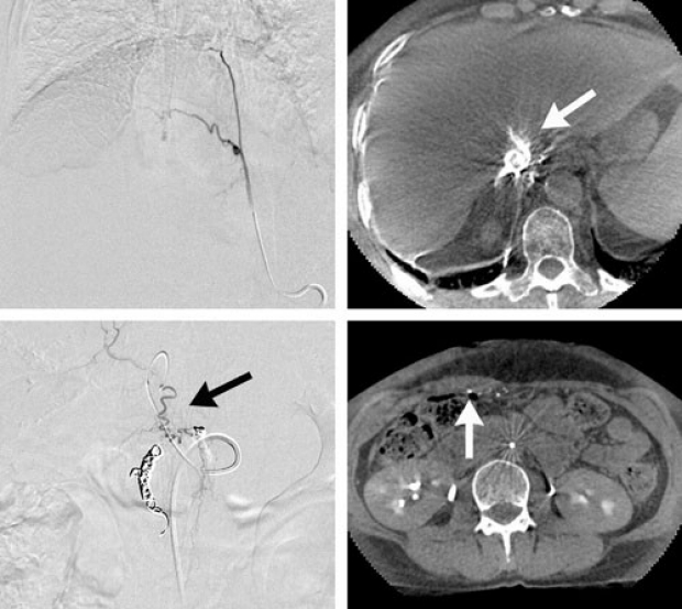
RESULTS: In 22 of 42 cases (52%), extrahepatic enhancement or incomplete tumor perfusion seen on CBCT affected the treatment plan. In 14 patients (33%), the findings were evident exclusively on CBCT and not detected by DSA. When comparing CBCT versus (99m)Tc MAA scintigraphy, CBCT showed eight cases of extrahepatic enhancement (19%) that were not evident on (99m)Tc MAA imaging. CBCT findings directed the additional embolization of vessels or repositioning of the catheter for better contrast agent and microsphere distribution. One case of gastric ulcer from nontarget embolization caused by reader error was observed.
CONCLUSIONS: CBCT can provide additional information about tumor and tissue perfusion not currently detectable by DSA or (99m)Tc MAA imaging, which should optimize (90)Y microsphere delivery and reduce nontarget embolization.
Transjugular intrahepatic portosystemic shunt creation in a polycystic liver facilitated by hybrid cross-sectional/angiographic imaging.
Sze DY, Strobel N, Fahrig R, Moore T, Busque S, Frisoli JK.
Department of Radiology, Stanford University Medical Center, H-3646, Stanford, California 94305-5642, USA.
Polycystic liver disease (PCLD) has long been considered to represent a contraindication to transjugular intrahepatic portosystemic shunt (TIPS) creation, primarily because of the risk of hemorrhage. Three-dimensional (3D) navigation within the enlarged and potentially disorienting parenchyma can now be performed during the procedure with the development of C-arm cone-beam computed tomography, which relies on the same equipment already used for angiography. Such a hybrid 3D reconstruction-enabled angiography system was used for safe image guidance of a TIPS procedure in a patient with PCLD. This technology has the potential to expedite any image-guided procedure that requires 3D navigation.
Utility of C-arm CT in patients with hepatocellular carcinoma undergoing transhepatic arterial chemoembolization.
Tognolini A, Louie JD, Hwang GL, Hofmann LV, Sze DY, Kothary N.
Division of Interventional Radiology, Stanford University Medical Center, 300 Pasteur Dr, H3652, Stanford, CA 94305-5642, USA.
PURPOSE: To evaluate the utility of C-arm computed tomography (CT) on treatment algorithms in patients undergoing transhepatic arterial chemoembolization for hepatocellular carcinoma (HCC).
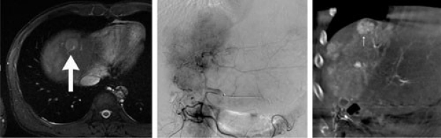
MATERIALS AND METHODS: From March 2008 to July 2008, 84 consecutive patients with HCC underwent 100 consecutive transhepatic arterial chemoembolizations with iodized oil. Unenhanced and iodinated contrast medium-enhanced C-arm CT with planar and three-dimensional imaging were performed in addition to conventional digital subtraction angiography (DSA) in all patients. The effect on diagnosis and treatment was determined by testing the hypotheses that C-arm CT, in comparison to DSA, provides (a) improved lesion detection, (b) expedient identification and mapping of arterial supply to a tumor, (c) improved characterization of a lesion to allow confident differentiation of HCC from pseudolesions such as arterioportal shunts, and (d) an improved evaluation of treatment completeness. The effect of C-arm CT was analyzed on the basis of information provided with C-arm CT that was not provided or readily apparent at DSA.
RESULTS: C-arm CT was technically successful in 93 of the 100 procedures (93%). C-arm CT provided information not apparent or discernible at DSA in 30 of the 84 patients (36%) and resulted in a change in diagnosis, treatment planning, or treatment delivery in 24 (28%). The additional information included, amongst others, visualization of additional or angiographically occult tumors in 13 of the 84 patients (15%) and identification of incomplete treatment in six (7.1%).
CONCLUSIONS: C-arm CT is a useful collaborative tool in patients undergoing transhepatic arterial chemoembolization and can affect patient care in more than one-fourth of patients.