General Applications
Dual-energy Imaging using an angiographic C-arm CT System

Mueller K, Datta S, Ahmad M, Choi J-H, Moore T, Pung L, Niebler C, Gold G E, Maier A, and Fahrig R.
Radiology, Stanford University, Lucas MRS Imaging Center 1201 Welch Rd., Palo Alto, California 94305, USA.
In the last years, dual-energy CT imaging has shown clinical value, thanks to its ability to differentiate materials based on their atomic number and to exploit different properties of images acquired at two different energies. C-arm CT systems are used to guide procedures in the interventional suite. Until now, there are no commercially available systems that employ dual-energy material decomposition. This paper explores the feasibility of implementing a fast kV-switching technique on a clinically available angiographic system for acquiring dual-energy C-arm CT images.
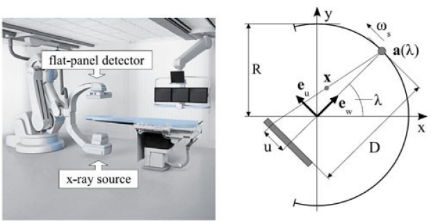
Dose and image quality for a cone-beam C-arm CT system.

Fahrig R, Dixon R, Payne T, Morin RL, Ganguly A, Strobel N.
Radiology, Stanford University, Lucas MRS Imaging Center 1201 Welch Rd., Rm. P-286, Palo Alto, California 94305, USA.
We assess dose and image quality of a state-of-the-art angiographic C-arm system (Axiom Artis dTA, Siemens Medical Solutions, Forchheim, Germany) for three-dimensional neuro-imaging at various dose levels and tube voltages and an associated measurement method. Unlike conventional CT, the beam length covers the entire phantom, hence, the concept of computed tomography dose index (CTDI) is not the metric of choice, and one can revert to conventional dosimetry methods by directly measuring the dose at various points using a small ion chamber. This method allows us to define and compute a new dose metric that is appropriate for a direct comparison with the familiar CTDIw of conventional CT. A perception study involving the CATPHAN 600 indicates that one can expect to see at least the 9 mm inset with 0.5% nominal contrast at the recommended head-scan dose (60 mGy) when using tube voltages ranging from 70 kVp to 125 kVp. When analyzing the impact of tube voltage on image quality at a fixed dose, we found that lower tube voltages gave improved low contrast detectability for small-diameter objects. The relationships between kVp, image noise, dose, and contrast perception are discussed.
A model for filtered backprojection reconstruction artifacts due to time-varying attenuation values in perfusion C-arm CT.
Fieselmann A, Dennerlein F, Deuerling-Zheng Y, Boese J, Fahrig R, Hornegger J.
Department of Computer Science, Pattern Recognition Lab, Friedrich-Alexander University of Erlangen-Nuremberg, Martensstr. 3, 91058 Erlangen, Germany.
Filtered backprojection is the basis for many CT reconstruction tasks. It assumes constant attenuation values of the object during the acquisition of the projection data. Reconstruction artifacts can arise if this assumption is violated. For example, contrast flow in perfusion imaging with C-arm CT systems, which have acquisition times of several seconds per C-arm rotation, can cause this violation. In this paper, we derived and validated a novel spatio-temporal model to describe these kinds of artifacts. The model separates the temporal dynamics due to contrast flow from the scan and reconstruction parameters. We introduced derivative-weighted point spread functions to describe the spatial spread of the artifacts. The model allows prediction of reconstruction artifacts for given temporal dynamics of the attenuation values. Furthermore, it can be used to systematically investigate the influence of different reconstruction parameters on the artifacts. We have shown that with optimized redundancy weighting function parameters the spatial spread of the artifacts around a typical arterial vessel can be reduced by about 70%. Finally, an inversion of our model could be used as the basis for novel dynamic reconstruction algorithms that further minimize these artifacts.
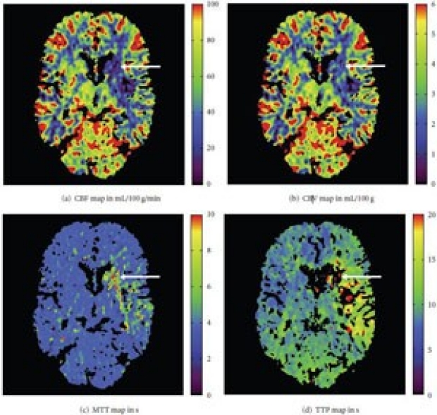
Deconvolution-Based CT and MR Brain Perfusion Measurement: Theoretical Model Revisited and Practical Implementation Details.
Fieselmann A, Kowarschik M, Ganguly A, Hornegger J, Fahrig R.
Pattern Recognition Lab, Department of Computer Science, Friedrich-Alexander University of Erlangen-Nuremberg, Martensstrafle 3, 91058 Erlangen, Germany.
Deconvolution-based analysis of CT and MR brain perfusion data is widely used in clinical practice and it is still a topic of ongoing research activities. In this paper, we present a comprehensive derivation and explanation of the underlying physiological model for intravascular tracer systems. We also discuss practical details that are needed to properly implement algorithms for perfusion analysis. Our description of the practical computer implementation is focused on the most frequently employed algebraic deconvolution methods based on the singular value decomposition. In particular, we further discuss the need for regularization in order to obtain physiologically reasonable results. We include an overview of relevant preprocessing steps and provide numerous references to the literature. We cover both CT and MR brain perfusion imaging in this paper because they share many common aspects. The combination of both the theoretical as well as the practical aspects of perfusion analysis explicitly emphasizes the simplifications to the underlying physiological model that are necessary in order to apply it to measured data acquired with current CT and MR scanners.
Dose and detectability for a cone-beam C-arm CT system revisited.

Ganguly A, Yoon S, Fahrig R.
Department of Radiology, Lucas MRS Center, Stanford University, 1201 Welch Road, Palo Alto, California 94305, USA.
PURPOSE: The authors had previously published measurements of the detectability of disk-shaped contrast objects in images obtained from a C-arm CT system. A simple approach based on Rose's criterion was used to scale the date, assuming the threshold for the smallest diameter detected should be inversely proportional to (dose)1/2. A more detailed analysis based on recent theoretical modeling of C-arm CT images is presented in this work.
METHODS: The signal and noise propagations in a C-arm based CT system have been formulated by other authors using cascaded systems analysis. They established a relationship between detectability and the noise equivalent quanta. Based on this model, the authors obtained a relation between x-ray dose and the diameter of the smallest disks detected. A closed form solution was established by assuming no rebinning and no resampling of data, with low additive noise and using a ramp filter. For the case when no such assumptions were made, a numerically calculated solution using previously reported imaging and reconstruction parameters was obtained. The detection probabilities for a range of dose and kVp values had been measured previously. These probabilities were normalized to a single dose of 56.6 mGy using the Rose-criteria-based relation to obtain a universal curve. Normalizations based on the new numerically calculated relationship were compared to the measured results.
RESULTS: The theoretical and numerical calculations have similar results and predict the detected diameter size to be inversely proportional to (dose)1/3 and (dose)1/2.8, respectively. The normalized experimental curves and the associated universal plot using the new relation were not significantly different from those obtained using the Rose-criterion-based normalization.
CONCLUSIONS: From numerical simulations, the authors found that the diameter of detected disks depends inversely on the cube root of the dose. For observer studies for disks larger than 4 mm, the cube root as well as square root relations appear to give similar results when used for normalization.
In-vitro imaging of femoral artery nitinol stents for deformation analysis

Ganguly A, Simons J, Schneider A, Keck B, Bennett NR, Fahrig R.
Department of Radiology, Stanford University, 1201 Welch Rd., Palo Alto, CA 94305, USA.
PURPOSE: Femoral artery stents are prone to fracture, and studying their deformations could lead to a better understanding of the cause of breakage. The present study sought to develop a method of imaging and analyzing stent deformation in vitro with use of a calibrated test device.
MATERIALS AND METHODS: High-resolution (approximately 200 μm) volumetric data were obtained with a flat-panel detector-based C-arm computed tomography system. A nitinol stent placed in a testing device was imaged with various loads that caused bending, axial tension, and torsion. Semiautomatic software was developed to calculate the bending, extension, and torsion from the stent images by measuring the changes in the radius of curvature, eccentricity, and angular distortions.
RESULTS: For the axial tension case, there was generally good agreement between the physical measurements and the image-based measurements. The bending measurements had better agreement at bend angles lower than 30∞. For stent torsion, the hysteresis between the loading and unloading curves were larger for the image-based results compared with physical measurements.
CONCLUSIONS: An imaging and analysis framework has been set up for the analysis of stent deformations that shows fairly good agreement between physical and image-based measurements.
An efficient estimation method for reducing the axial intensity drop in circular cone-beam CT.

Zhu L, Starman J, Fahrig R.
Department of Radiology, Stanford University, Stanford, CA 94305, USA.
Reconstruction algorithms for circular cone-beam (CB) scans have been extensively studied in the literature. Since insufficient data are measured, an exact reconstruction is impossible for such a geometry. If the reconstruction algorithm assumes zeros for the missing data, such as the standard FDK algorithm, a major type of resulting CB artifacts is the intensity drop along the axial direction. Many algorithms have been proposed to improve image quality when faced with this problem of data missing; however, development of an effective and computationally efficient algorithm remains a major challenge. In this work, we propose a novel method for estimating the unmeasured data and reducing the intensity drop artifacts. Each CB projection is analyzed in the Radon space via Grangeat's first derivative. Assuming the CB projection is taken from a parallel beam geometry, we extract those data that reside in the unmeasured region of the Radon space. These data are then used as in a parallel beam geometry to calculate a correction term, which is added together with Hu's correction term to the FDK result to form a final reconstruction. More approximations are then made on the calculation of the additional term, and the final formula is implemented very efficiently. The algorithm performance is evaluated using computer simulations on analytical phantoms. The reconstruction comparison with results using other existing algorithms shows that the proposed algorithm achieves a superior performance on the reduction of axial intensity drop artifacts with a high computation efficiency.